2024_06_18 Neurocysticercosis, Annotated
Long overdue correction: I know now that this first pair of images (Midbrain/spinal cord) depicts blood vessels; hence the strikethroughs. Re: the Sagittal view, I read an article saying that particular artifact is new as of 3T MRI technology. It galls to think the others might be something, and because I prefaced them with a mistake, they might have lost impact. -2024_12_13
Neurocysticercosis suspects
To respect your time, I’ll only share the clearest examples.
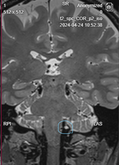
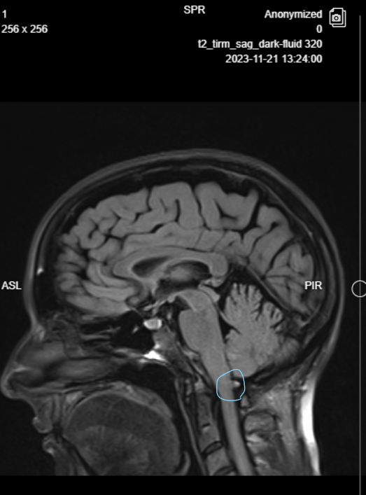
Midbrain/spinal cord
The blip on coronal view here can be appreciated over multiple frames, many which show possible centrally adjacent edema. The second blip unfortunately only exists in this sagittal frame… couldn’t be an artifact?
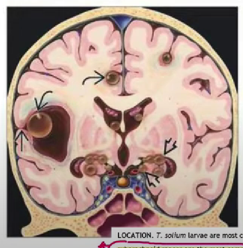
Basal ganglia level
If you look at enough examples of Neurocysticercosis imaging, you’ll note that the pathognomonic “cyst with dot” sign is not really just a single eccentric dot, and can often be observed as a distinctive big dot/ little dot pair.
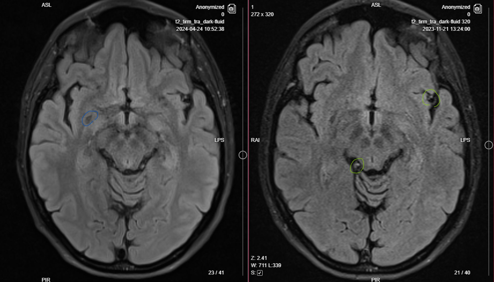
[I suspect the 5mm VR space we talked about is a cysticercus because 1.Two dots, and 2. (appearance of) non-CSF intensity (cysticerci in the same brain can be at different stages of breakdown).]
The two in yellow circles are of course the better candidates.
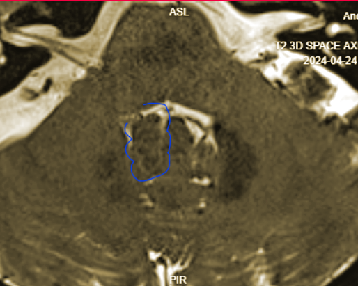
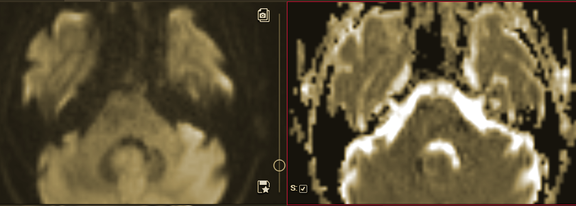
4th ventricle
Racemose cystic grouping? Restricted diffusion?
Related:
“Large subarachnoid cysts often adopt a multilobulated appearance due to the confluence of vesicles together with inflamed arachnoid membranes; this imaging resembling a “bunch of grapes” is highly suggestive of NCC” (https://doi.org/10.1016/j.jns.2016.11.045 Del Brutto et al)
Comments
Post a Comment